Radiology and CT
The Bone and Joint Imaging and Intervention Program at Narmada Health Group, Imaging offers state-of-the-art diagnostic exams including Spiral CT, Digital X-ray, USG, PFT and TMT interpretation by specialty-trained radiologists, and image-guided procedures for the bones, joints, and spine. At Narmada Imaging, every scan is read by a specialty-trained radiologist: an expert who has extensive training and real-world experience diagnosing - experts in the spine, bones, joints and muscles. This group includes recognized authorities on sports-related injuries, bone and muscle tumors, and minimally invasive, image-guided treatments for joint and spine disorders. Members of the team have helped to develop state-of-the-art tests and treatments for conditions such as joint pain and osteoporosis.
Our Radiology Center has :
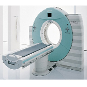
- Spiral CT-Scan
- High frequency S-ray, 500mA (Siemens), 60mA and 30mA X-Ray machines
- CT-Scan
- Digital X-ray
- USG
- PFT, TMT
- ECHO
- Color Doppler